This is the second of three posts on the 2015 European Science Photo Competition. We published the first in December 2015 and the third in March 2016.
The 2015 European Science Photo Competition saw 9793 files uploaded from 2201 authors from 40 countries. Naturally, many of them didn’t make it into the final, but there were lot of great shots besides the 328 submissions (384 separate images[1]) that went on to compete for first place.
All of the files in this competition were freely licensed under CC BY-SA 4.0, including the following:
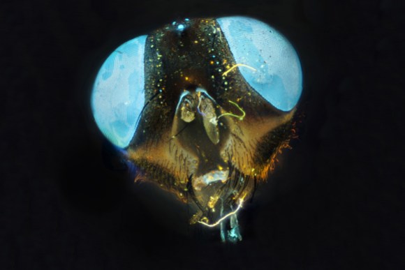
Fly under fluorescence microscope. Pre-coloring with acridine orange, luminescence excitation by ultraviolet light. Photo by Alexander Klepnev, Russia.
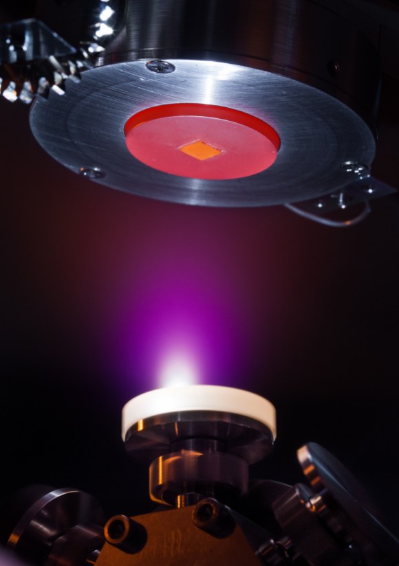
Thin films of oxides are deposited with atomic layer precision using pulsed laser deposition. A high-intensity pulsed laser is shooting onto the rotating white target consisting of Al2O3 (alumina). The laser pulse creates a plasma explosion visible as the purple cloud. The plasma cloud expands towards the square substrate, consisting of SrTiO3, where it deposits thin layers of alumina, one atomic layer at a time. This results in a conducting interface between the two materials which are otherwise insulating. The substrate is mounted on a heating plate, glowing red from a temperature of 650 °C, to improve the crystallinity of the alumina thin film. Photo by Adam Andersen Læssøe, Denmark.
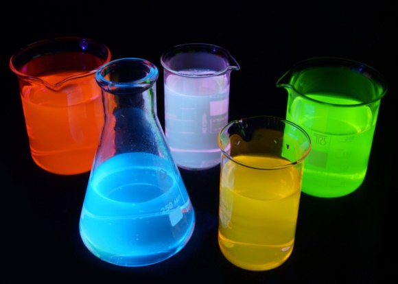
In this picture you can see fluorescence of different substances under UV light. Green is a fluorescein, red is rhodamine B, yellow is rhodamine 6G, blue is quinine, purple is mixture of quinine and rhodamine 6G. Solutions are about 0,001% concentration in water. Photo by Maxim Bilovitskiy, Estonia.
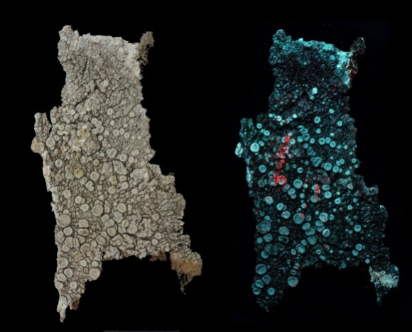
Same lichen under normal light and ultraviolet light. Photo by Maria Milaslava, Russia.
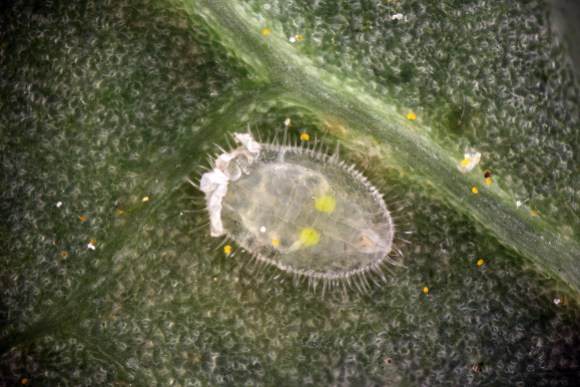
Greenhouse whitefly (Trialeurodes vaporariorum) larva. Light microscopy, incident light. Sharpness is achieved by focus stacking. Photo by Anatoly Mikhaltsov, Russia.
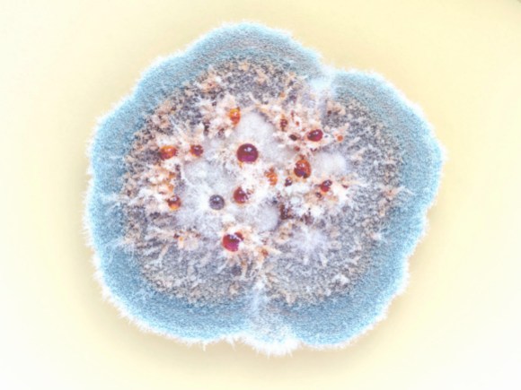
A colony of Penicillium sp. in Petri dish. Photo by Milo Manica, Italy.
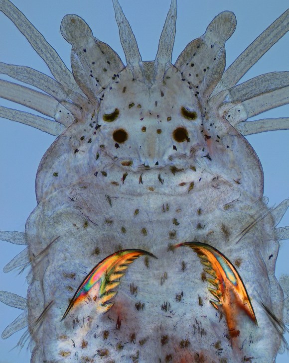
Polychaete errantia marine worm. 10x magnification under polarized light. Photo by Cosmic73, Greece.
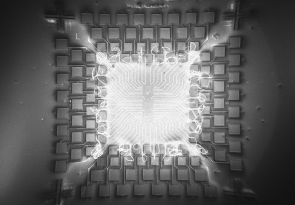
SEM image of carbon nanotube based field-effect transistor in fabrication. Image obtained during one of the fabrication steps, after hydrofluoric acid wet etching and SF6 based inductively coupled plasma dry etching steps. Glowing structures in the middle are charged photoresist residues. In this chip design each square test pad represents transistor’s source or drain electrodes, across which single-walled carbon nanotube will be suspended during the subsequent fabrication stages. Scale: the width of the outermost square test pads is 150 micrometers. SEM image and fabrication by Aidar Kemelbay (NLA, Nazarbayev University, Kazakhstan), with the help of Dr. Emine Cagin (NTB Buchs, Switzerland).
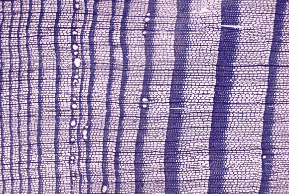
Cross section of Larix gmelinii, growing in the north of Siberia. Photo by Maria Tabakova, Russia.
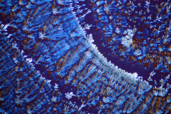
Microcrystals of mineral water “Essentuki 4”. Light microscopy with polarized light. Micrograph by Dashko Alexandra, student of The Science Research Laboratory “Microcosmos”. Photo by Mihaltsov Anatoly Ivanovich, Russia.
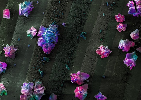
Indium phosphide nanocrystalline surface obtained by electrochemical etching. False colours. Photos of nanostructures were obtained in scanning electron microscope JSM-6490 by the researchers of Berdyansk State Pedagogical University. Photo by Яна Сычикова and Сергей Ковачев, Ukraine.
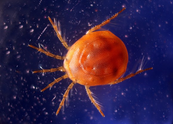
Water mite. This animal has length of 1.1 mm. The water sample is taken from a shallow freshwater pond. Light microscopy, incident light, focus stacking. Photo by Anatoly Mikhaltsov, Russia.
Ivo Kruusamägi, Estonian Wikipedian, European Science Photo Competition organizer

Can you help us translate this article?
In order for this article to reach as many people as possible we would like your help. Can you translate this article to get the message out?
Start translation